
Page 90 - 14_Atti_SIN_SNO_2022_flip
P. 90
Proceedings SNO “Percorsi clinici in Neuroscienze”
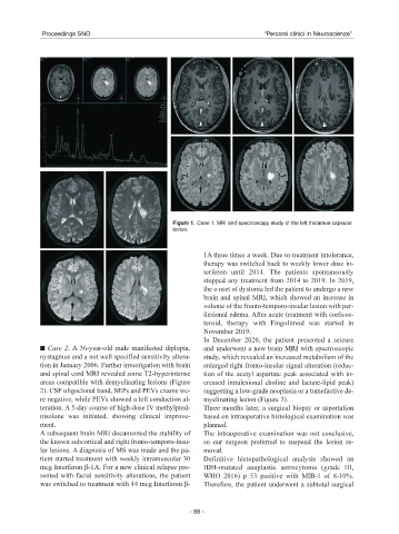
Figure 1. Case 1. MRI and spectroscopy study of the left thalamus-capsular
lesion.
1A three times a week. Due to treatment intolerance,
therapy was switched back to weekly lower dose in-
terferon until 2014. The patients spontaneously
stopped any treatment from 2014 to 2019. In 2019,
the o nset of dystonia led the patient to undergo a new
brain and spinal MRI, which showed an increase in
volume of the fronto-temporo-insular lesion with per-
ilesional edema. After acute treatment with corticos-
teroid, therapy with Fingolimod was started in
November 2019.
In December 2020, the patient presented a seizure
■ ■ Case 2. A 36-year-old male manifested diplopia, and underwent a new brain MRI with spectroscopic
nystagmus and a not well specified sensitivity altera- study, which revealed an increased metabolism of the
tion in January 2006. Further investigation with brain enlarged right fronto-insular signal alteration (reduc-
and spinal cord MRI revealed some T2-hyperintense tion of the acetyl aspartate peak associated with in-
areas compatible with demyelinating lesions (Figure creased intralesional choline and lactate-lipid peak)
2). CSF oligoclonal band, SEPs and PEVs exams we- suggesting a low-grade neoplasia or a tumefactive de-
re negative, while PEVs showed a left conduction al- myelinating lesion (Figure 3).
teration. A 5-day course of high-dose IV methylpred- Three months later, a surgical biopsy or asportation
nisolone was initiated, showing clinical improve- based on intraoperative histological examination was
ment. planned.
A subsequent brain MRI documented the stability of The intraoperative examination was not conclusive,
the known subcortical and right fronto-temporo-insu- so our surgeon preferred to suspend the lesion re-
lar lesions. A diagnosis of MS was made and the pa- moval.
tient started treatment with weekly intramuscular 30 Definitive histopathological analysis showed an
mcg Interferon β-1A. For a new clinical relapse pre- IDH-mutated anaplastic astrocytoma (grade III,
sented with facial sensitivity alterations, the patient WHO 2016) p 53 positive with MIB-1 of 8-10%.
was switched to treatment with 44 mcg Interferon β- Therefore, the patient underwent a subtotal surgical
- 88 -