
Page 91 - 14_Atti_SIN_SNO_2022_flip
P. 91
Proceedings SNO “Percorsi clinici in Neuroscienze”
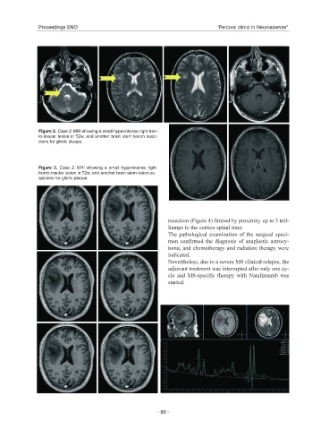
Figure 2. Case 2. MRI showing a small hyperintense right fron-
to-insular lesion in T2w, and another brain stem lesion suspi-
cions for gliotic plaque.
Figure 3. Case 2. MRI showing a small hyperintense right
fronto-insular lesion in T2w, and another brain stem lesion su-
spicions for gliotic plaque.
resection (Figure 4) limited by proximity up to 3 mil-
liamps to the cortico spinal tract.
The pathological examination of the surgical speci-
men confirmed the diagnosis of anaplastic astrocy-
toma, and chemotherapy and radiation therapy were
indicated.
Nevertheless, due to a severe MS clinical relapse, the
adjuvant treatment was interrupted after only one cy-
cle and MS-specific therapy with Natalizuamb was
started.
- 89 -