
Page 92 - 14_Atti_SIN_SNO_2022_flip
P. 92
Proceedings SNO “Percorsi clinici in Neuroscienze”
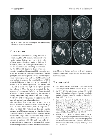
Figure 4. Case 2. Pre- and post-surgical MRI demonstrating
the subtotal removal of the lesion.
DISCUSSION
As other works pointed out , many intracranial
(3,4)
conditions, like CNS tumors, can present with
white matter lesions and can mimic MS.
Clinical presentation is not useful in differential
diagnosis, as well as radiological features which
do not reach sufficient sensitivity and specifici-
ty for differential diagnosis. Even when con-
fronting a confirmed diagnosis of MS, atypical symp- cern. However, further analyses with more compre-
toms, or uncommon radiological evolution, should hensive cohorts and prospective studies are needed to
prompt further investigations. Based on our experi- support our data.
ence, we retrospectively analyzed the MS database of
our institute to evaluate the exact incidence of CNS
tumor in MS patients from 2011 to 2021. Overall, 15 REFERENCES
out of 3010 MS patients presented CNS tumors, and
7 out of 15 cases were high- or low-grade gliomas 1. Oh J, Vidal-Jordana A, Montalban X. Multiple sclerosis:
(prevalence 0,49%). We also investigated the fre- clinical aspects. Curr Opin Neurol 2018; 31 (6): 752-759.
quency of post-surgical infection or hematological 2. Lin X, Yu WY, Liauw L, Chander RJ, Soon WE, Lee HY,
disorders in those patients receiving immunomodu- Tan K. Clinicoradiologic features distinguish tumefactive
lating therapy. Our study revealed that none of the pa- multiple sclerosis from CNS neoplasms. Neurol Clin
tients who underwent surgical procedures presented Pract 2017; 7 (1): 53-64.
any relevant perisurgical complications. 3. Geraldes R, Ciccarelli O, Barkhof F, De Stefano N,
Our experience demonstrates that in some cases, a Enzinger C, Filippi M, Hofer M, Paul F, Preziosa P,
careful evaluation is needed in the differential diag- Rovira A, DeLuca GC, Kappos L, Yousry T, Fazekas F,
Frederiksen J, Gasperini C, Sastre-Garriga J, Evangelou
nosis of white matter lesions, as CNS neoplastic le-
N, Palace J; MAGNIMS study group. The current role of
sions can mimic MS. Furthermore, they can occur in
MRI in differentiating multiple sclerosis from its imaging
patients with MS presenting new or enlarging lesions. mimics. Nat Rev Neurol 2018; 14 (4): 199-213.
Surgical biopsy is often needed for diagnostic confir-
4. Kim DS, Na DG, Kim KH, Kim JH, Kim E, Yun BL,
mation. Our single center retrospective study demon-
Chang KH. Distinguishing tumefactive demyelinating le-
strates that the incidence of CNS tumors is not in-
sions from glioma or central nervous system lymphoma:
creased in patients with MS, and even under im- added value of unenhanced CT compared with conven-
munomodulating treatment, a neurosurgical proce- tional contrast-enhanced MR imaging. Radiology. 2009;
dure can be performed without additional safety con- 251 (2): 467-475.
- 90 -