
Page 78 - 14_Atti_SIN_SNO_2022_flip
P. 78
Proceedings SNO “Percorsi clinici in Neuroscienze”
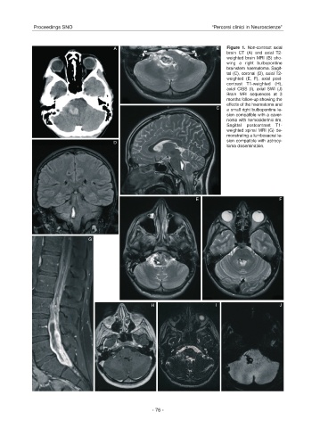
A B Figure 1. Non-contrast axial
brain CT (A) and axial T2-
weighted brain MRI (B) sho-
wing a right bulbopontine
brainstem haematoma. Sagit-
tal (C), coronal (D), axial T2-
weighted (E, F), axial post-
contrast T1-weighted (H),
axial CISS (I), axial SWI (J)
Brain MRI sequences at 3
months follow-up showing the
effects of the haematoma and
C a small right bulbopontine le-
sion compatible with a caver-
noma with hemosiderinic rim.
Sagittal postcontrast T1-
weighted spinal MRI (G) de-
monstrating a lumbosacral le-
D sion compatible with astrocy-
toma dissemination.
E F
G
H I J
- 76 -